
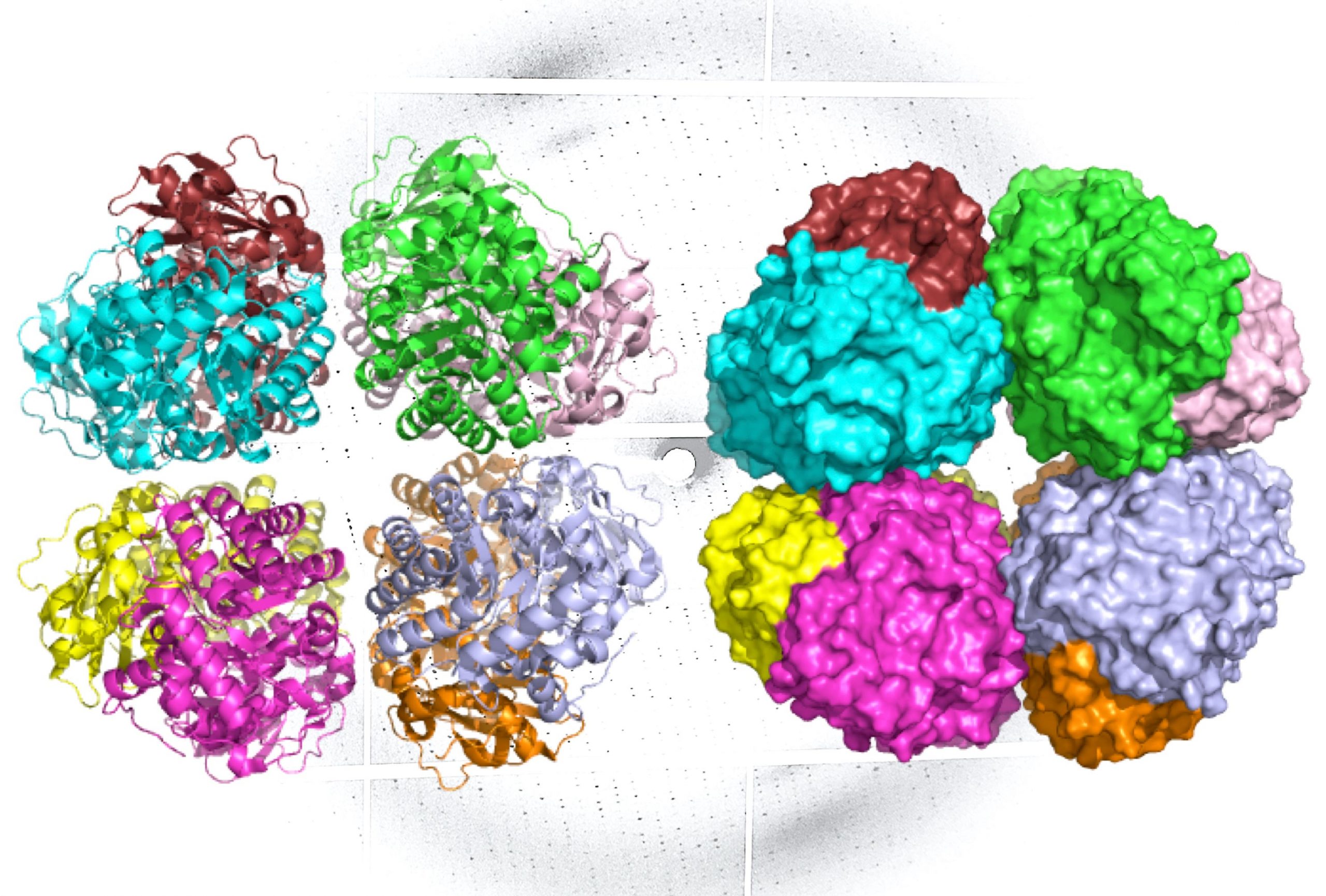
A band diagram (L) and a representation of the molecular surface area (R) of the carbon-fixing form I ‘rubisco show eight molecular subunits without the small subunits. In the background there is an X-ray diffraction pattern of the enzyme, which was also generated by the research team. Photo credit: Henrique Pereira / Berkeley Lab
A team of scientists have discovered an ancient form of Rubisco, the most abundant enzyme on earth that is vital to life as we know it.
The newly identified Rubisco was found in previously unknown environmental microbes and offers insights into the evolution of the photosynthetic organisms that underlie the planet’s food chains.
“Rubisco is the main driver for the production of food, so it can absorb CO2 from the atmosphere and fix it in sugar so that plants and other photosynthetic organisms can use it, ”said Doug Banda, postdoctoral fellow in Patrick Shih’s lab, assistant professor at UC Davis and director of Plant Biosystems Design at the Joint BioEnergy Institute (JBEI) administered by the Lawrence Berkeley National Laboratory (Berkeley Lab). “It’s also one of the oldest carbon-fixing enzymes on the planet.”
Form I Rubisco, which is found in plants, algae, and cyanobacteria, has a deep evolutionary history with the planet that goes back nearly 2.4 billion years to the great oxygenation event when cyanobacteria literally transformed the earth’s atmosphere by bringing in oxygen through photosynthesis. Rubisco’s role in this landmark event makes it a focal point for scientists studying the evolution of life as well as scientists looking to develop bio-based fuels and renewable energy technologies.
One study appears in Natural plants, Banda, and researchers at UC Davis, UC Berkeley, and Berkeley Lab report the discovery and characterization of a previously undescribed lineage of Form I Rubisco – one that researchers suspect may have deviated from Form I Rubisco prior to the development of cyanobacteria.
The new line, called Form I ‘Rubisco, was found through metagenomic analysis of environmental samples and synthesized in a laboratory. It gives researchers new insights into the structural evolution of Form I Rubisco and possibly provides clues as to how this enzyme changed the planet.
“This is what a Rubisco could have looked like before oxygen rose more than 2.4 billion years ago,” Shih said, noting that the Shape I ‘Rubisco offers scientists a window on how ancient microbes might have fixed carbon from the rise of carbon Cyanobacteria and the Form I Rubisco.
An invisible world
Form I Rubisco is a hexadecamer, which means it consists of eight large molecular subunits with eight small subunits on top and bottom. Every piece of the structure of this protein is an essential part of photosynthesis and therefore the carbon fixation process.
Other functional forms of Rubisco exist in bacteria and microorganisms of the archaeal domain. These variants come in different shapes and sizes, and they all perform the same step of photosynthesis. However, Form I Rubisco is responsible for the vast majority of carbon sequestration on Earth.
Study co-author and contributor, Professor Jill Banfield of UC Berkeley’s Department of Earth and Planetary Sciences, discovered Form I ‘Rubisco after performing metagenomic analyzes on groundwater samples. Metagenomic analyzes allow researchers to study genes and genetic sequences of uncultivated microorganisms in the environment.
Using the genes and genetic sequences provided by Banfield, Banda and Shih, the form of I’-Rubisco has been successfully expressed in the laboratory using E. coli. To learn how this newly identified shape works and how it compares to previously discovered Rubisco enzymes, the scientists had to create precise 3D models of its structure. For this task, the lead authors turned to structural biologists Paul Adams, Henrique Pereira, and Michal Hammel from the Berkeley Lab.
First, Adams and Pereira performed X-ray crystallography at Berkeley Lab’s Advanced Light Source (ALS) – an approach that can produce images of molecules with atomic resolution. To understand how the structure of the enzyme changes during different states of activity, Hammel applied a technique called small angle X-ray scattering (SAXS), using the SIBYLS beamline at the ALS.
SAXS is a lower resolution technique, but unlike crystallography, which requires sample molecules to be frozen in crystal form, SAXS is performed in solution. When the data from the two approaches are combined, scientists can construct unprecedented models of complex molecules as found in nature.
“Like many enzymes that are vital to life, Rubisco has multiple protein domains linked together, and because it binds to other molecules during the photosynthetic reaction, it goes through various arrangements of those domains,” said Hammel, biophysicist at Berkeley Lab’s Molecular Biophysics and Integrated Bioimaging Department (MBIB). “Our techniques really worked hand in hand to show how this new, novel Rubisco would behave under real physiological conditions.”
The ALS investigations showed that Form I ‘Rubisco, like Form I Rubisco, is made up of eight large subunits. However, it does not possess the small subunits previously believed to be essential for its carbon-fixing function.
The researchers now believe that Form I ‘Rubisco is a missing link in the evolutionary history of Form I Rubisco’s structure.
“The discovery of an octameric rubisco that forms without small subunits enables us to ask [evolutionary] Asking what life would have been like without the functionality of small subunits, ”said Banda.
Following the success of the structural study of Form I ‘Rubisco, Shih commissioned Hammel, Adams, and Pereira to apply their complementary approach to studies of other key plant enzymes, including additional forms of Rubisco.
“We have worked together at the Berkeley Lab for over 10 years and it has been really satisfying to see what crystallography and SAXS can do together to understand biological problems,” said Pereira, an MBIB biophysicist. “In the past, the scientists who use these various structural biology techniques would have seen each other as being in competition and would have tried each other to solve structures. But now it’s pure collaboration. ”
For more information on this research, see Missing Link Discovered in the Evolution of Photosynthesis and Carbon Fixation.
Reference: “Novel bacterial clade reveals the origin of Form I Rubisco” by Douglas M. Banda, Jose H. Pereira, Albert K. Liu, Douglas J. Orr, Michal Hammel, Christine He, Martin AJ Parry, Elizabete Carmo-Silva, Paul D. . Adams, Jillian F. Banfield, and Patrick M. Shih, Aug 31, 2020; Natural plants.
DOI: 10.1038 / s41477-020-00762-4
The ALS is a user facility of the Ministry of Energy (DOE) and the JBEI is a DOE bioenergy research center. The crystallographic beamline used in this research is operated by the Berkeley Center for Structural Biology and funded by the Howard Hughes Medical Institute. The SIBYLS beamline is supported by the National Cancer Institute Grant Structural Biology of DNA Repair and the DOE Office of Science. This work was partially supported by the DOE Office of Science.