
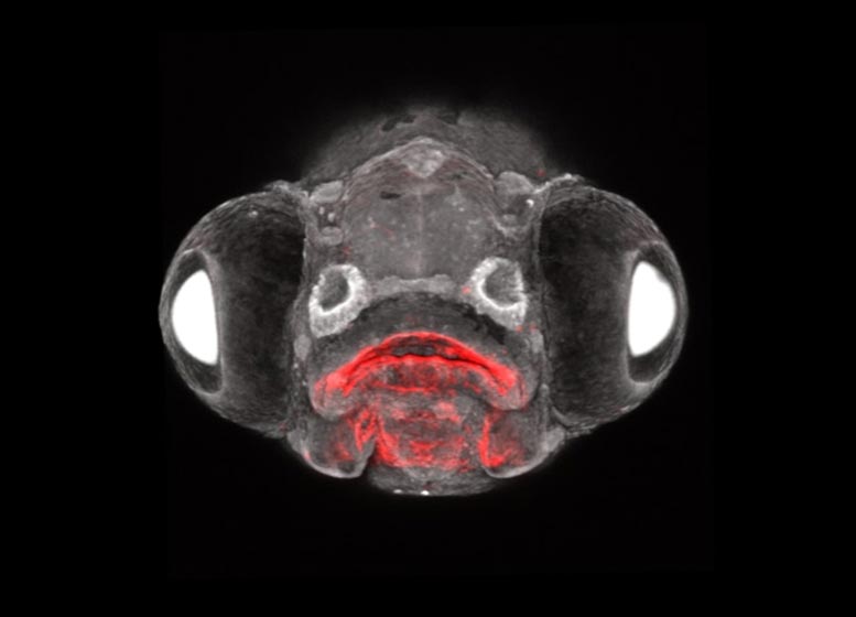
The view of the zebrafish face of the larvae shows a type of endoderm cell in red and all other cell types in white. Fabian, Crump, and colleagues used these transgenic zebrafish to show that endoderm cells make unexpected contributions to the pituitary gland, solving a long-standing mystery of pituitary development. Photo credit: Peter Fabian and Gage Crump / USC Stem Cell
Insights into centuries-old controversies over the development of the key gland come from research by the Keck School of Medicine of USC
A new USC-led study suggests a change in the developmental and evolutionary history of the pituitary gland.
The pea-sized gland located at the base of the brain produces hormones that promote growth, aggression, sexual development, and reproduction. For decades it was thought that the anterior lobe of the pituitary gland – where the hormones are made – was an evolutionary development that occurred in vertebrates along with the ear, nose and lens of the eye.
The widely accepted “new head hypothesis” is that all of these body parts come from some type of embryonic structure that is located in the ectoderm, or the outermost layer of an embryo. Meanwhile, animals with a spinal cord but without a backbone, believed to represent an earlier evolutionary step, have a pituitary-like structure that was previously believed to have a particular origin in the innermost embryonic layer or endoderm .

Gage Crump is a professor of stem cells and regenerative medicine at USC’s Keck School of Medicine. Photo credit: Richard Carrasco
In an article published on October 23, 2020 in scienceUSC researchers present evidence that in some vertebrates, the endoderm is also part of the anterior lobe of the pituitary gland – an idea that has been the subject of scientific controversy for more than 100 years. The results of the study, which was supported by a large grant from the National Institutes of Health, suggest that the gland may have a longer history of development than previously thought.
“We have repeated very ancient observations using cutting-edge technology that prove this idea that there is an endodermal contribution to the pituitary gland,” said senior author Gage Crump, professor of stem cell and regenerative medicine at USC’s Keck School of Medicine. “Our work revises ideas about what type of embryonic structure the pituitary gland is and when it first developed.”
Development detective work
Crump and colleagues studied zebrafish, a species that is useful as a laboratory model in part because its development is an open book to researchers. Eggs are fertilized from the outside and embryos are almost transparent. The research team used new laboratory methods of their own invention to mark the zebrafish’s embryonic cells and follow the cells that descended from them in adulthood.
In addition, they used time-lapse photography with a powerful microscope and single cells RNA Sequencing. This latter technology is related to DNA Sequencing, but rather than characterizing the entire genetic code, it only shows the genes that are simultaneously expressed for each of thousands of cells and in what amount – a powerful way to understand the nature of the cells under study.
In a series of time-lapse experiments using zebrafish embryos, the team documented Rathke’s pouch, a structure made up of the outer layer previously believed to be the sole source of the anterior lobe of the pituitary gland, and merged with Seessel’s pouch, a structure made up of the inner layer. Their observations show that the endoderm was responsible for about 20 percent of the cells in the anterior lobe of the pituitary gland.
Another experiment that followed the fate of embryonic cells in adult zebrafish showed a mixture of ectodermal and endodermal cells in the pituitary gland. When studying gene expression using RNA sequencing, the scientists found that cells from the inner endoderm layer differentiated into all of the main types of hormone-producing cells in the pituitary gland. In addition, in genetically engineered zebrafish embryos lacking the ectodermal component, endodermal cells may themselves form a pituitary-like structure, albeit much smaller than the normal pituitary.
Taken together, these studies clearly show an endodermal contribution to the zebrafish pituitary. This unexpected revelation suggests that the quasi-pituitary seen in certain more primitive non-vertebrates – underwater creatures that are weird and largely dark – may have survived in some form among at least some of their spinal evolutionary offspring.
Crump, who is also the founding director of USC’s Development, Stem Cells and Regenerative Medicine program, warns that it remains to be seen whether the residue will persist in humans.
“Fish may keep this ancestral trait, but humans have lost it,” he said. “We can see that the pituitary gland is not a brand new vertebrate structure like the nose, ear or lens, but existed before vertebrates and subsequently absorbed new ectoderm contributions. By capturing this evolutionary relic in the zebrafish, we have solved the mystery of where the pituitary gland came from. ”
An old idea that (surprisingly) came back
As early as the mid-1910s, anatomists had reported that Rathke’s pouch was closely connected to Seessel’s pouch. Rathke’s pouch has long been known as the source of the endocrine component of the pituitary gland. In contrast, the fate and purpose of Seessel’s pouch has remained a mystery to this day.
For researchers in the early 20th century (and more or less since then) there weren’t any good ways to further study the relationship between the two embryonic structures, and thus the possibility of an endodermal contribution to the pituitary gland. Initially hotly debated, the topic eventually turned into a historical curiosity.
Fortunately, lead author Peter Fabian, a postdoctoral fellow at the Keck School, was very familiar with this precedent. While trying one of these new embryonic cell tagging and tracking techniques developed by staff at the Sanford Burnham Prebys Medical Discovery Institute, he noticed endodermal cells in the pituitary gland. A lightbulb went out in his head.
“It was an accidental discovery,” said Crump. “We’re interested in the endoderm in general, but we didn’t set out to examine the pituitary gland. Since this was such an unexpected observation, we really had to prove it with multiple lines of investigation. ”
Reference: “Line analysis shows an endodermal contribution to the pituitary gland of vertebrates” by Peter Fabian, Kuo-Chang Tseng, Joanna Smeeton, Joseph J. Lancman, P. Duc Si Dong, Robert Cerny and J. Gage Crump, October 23, 2020, science.
DOI: 10.1126 / science.aba4767
Other authors on the study include Kuo-Chang Tseng of the Keck School; former Keck School Postdoc Joanna Smeeton, now from Columbia University;; Joseph J. Lancman and P. Duc Si Dong of Sanford Burnham Prebys; and Robert Cerny from Charles University in Prague.
This study was supported by the NIH (R35DE027550, K99DE027218, U01DK105541, DP2DK098092), the WM Keck Foundation, and the Czech Science Foundation (19-18634S).

