
Kaplan-Meier curves for all-cause mortality in patients with versus without myocardial injury (Panel A) and in patients with versus without myocardial injury for the presence or absence of serious echocardiographic abnormalities (Panel B). * Includes wall movement disorders, global left ventricular dysfunction, diastolic dysfunction, right ventricular dysfunction, and the presence of pericardial effusion. Event rates are censored 20 days after hospital admission. Photo credit: Mount Sinai Health System
Cardiac ultrasound (also known as echocardiograms) provides a look at the heart and the effects of the COVID-19 Virus in patients.
A new study by researchers at Mount Sinai’s Icahn School of Medicine identifies several types of cardiac structural damage that occur in COVID-19 patients after a heart injury and that can be linked to fatal conditions such as heart attack, pulmonary embolism, heart failure, and myocarditis. These abnormalities are associated with a higher risk of death in hospital patients. The results, published in the October 26, 2020 edition of Journal of the American College of Cardiologyprovide new insights that can help clinicians better understand the mechanism of heart injury, leading to faster identification of at-risk patients and guidance for future therapies.
“Detecting structural abnormalities early may require more appropriate treatments, including anticoagulation and other approaches, for patients in and out of the hospital,” says Dr. med. Valentin Fuster, Director of Mount Sinai Heart and Chief Physician of Mount Sinai Hospital.
The international, retrospective study extends previous Mount Sinai research showing that myocardial (heart damage) injuries are widespread in hospitalized patients with COVID-19 and associated with a higher risk of mortality. This study focused on patients’ troponin levels – proteins released when the heart muscle is damaged – and their findings (higher troponin levels mean greater heart damage).
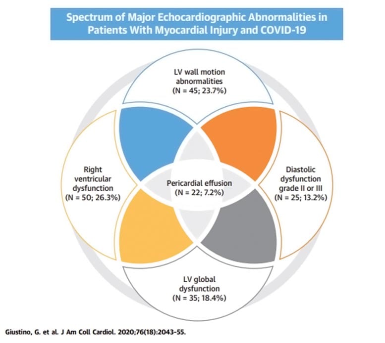
In patients with Covid-19 who had a TTE, almost two-thirds of patients with myocardial injury developed cardiac structure disorders. Cardiac structural abnormalities included right ventricular dysfunction, left ventricular wall movement disorders, global left ventricular dysfunction, diastolic dysfunction, and pericardial effusions. LV = left ventricular. Photo credit: Mount Sinai Health System
This new work examined the presence of cardiac troponin elevations in combination with the presence of echocardiographic abnormalities and found that the combination was associated with worse prognosis and mortality than troponin elevations alone.
“This is one of the first studies to provide detailed echocardiographic and electrocardiographic data in hospitalized patients with COVID-19 and laboratory evidence of myocardial injuries,” explains the first and corresponding author, Dr. med. Gennaro Giustino, Cardiology Fellow at Mount Sinai Hospital. “We found that in COVID-19 patients who had transthoracic echocardiography, these cardiac structural abnormalities were diverse and occurred in nearly two-thirds of the patients.”
The researchers examined transthoracic echocardiographic (TTE) and electrocardiographic (EKG) scans of 305 adult patients with confirmed positive COVID-19 who were admitted to four New York Mount Sinai health care system hospitals (Mount Sinai Hospital, Mount Sinai West, Mount Sinai Queens) and Mount Sinai (Beth Israel), Elmhurst Hospital in Queens and two hospitals in Milan, Italy between March and May 2020. The median age was 63 years and 67.2 percent were men. 190 patients (62.6 percent) had evidence of myocardial injury; 118 of them had heart damage at the time of hospitalization and 72 developed myocardial injury during hospitalization. The researchers found that patients with myocardial injury had more electrocardiographic abnormalities, higher inflammatory biomarkers, and an increased prevalence of TTE abnormalities compared to patients without heart injury.
The abnormalities were varied, with some patients exhibiting multiple abnormalities. 26.3 percent had right ventricular dysfunction (which may be associated with pulmonary embolism and severe respiratory failure), 23.7 percent had regional movement disorders of the left ventricular wall (which may be associated with heart attacks), 18.4 percent had diffuse left ventricular dysfunction (which 13.2 percent had grade II or III diastolic dysfunction (a condition that resulted in stiffer chambers of the heart), and 7.2 percent had pericardial effusions (extra fluid around the heart that causes the heart to pumping abnormally).
The study looked at hospital mortality and troponin elevation. It shows that the troponin elevation in patients without heart injury was 5.2 percent, compared with 18.6 percent in patients with myocardial injury but no echocardiographic abnormalities and 31.7 percent in patients with myocardial injury who also had echocardiographic abnormalities. The researchers cleared up for other major complications of COVID-19, including shock, acute respiratory distress syndrome, and kidney failure.
“Our study shows that an echocardiogram, performed with adequate personal protection, is a useful and important tool in the early detection of patients at higher risk of COVID-19 related heart injuries who may be from a more aggressive therapeutic approach early in their hospitalization benefit. Says the author in question, Dr. med. Martin Goldman, Dr. Arthur M. and Dr. Hilda A. Master Professor of Medicine (Cardiology) at the Icahn School of Medicine on Mount Sinai. “As this is a new disease with persistent symptoms, we also plan to follow these patients closely with imaging to evaluate the development and hopefully resolution of these heart problems.”
“Echocardiograms have been invaluable in providing critical information about patients with multiple heart conditions. Echocardiography is the only imaging method that can be brought to bed and used safely for patients, including those on ventilators, ”says Dr. Lori Croft, Associate Professor of Medicine (Cardiology) at the Icahn School of Medicine at Mount Sinai and Director of the Echocardiography Laboratory at Mount Sinai Hospital. “Our results will help to control the supply of Covid-19 patients at a critical time.”
Reference: October 26, 2020, Journal of the American College of Cardiology.