
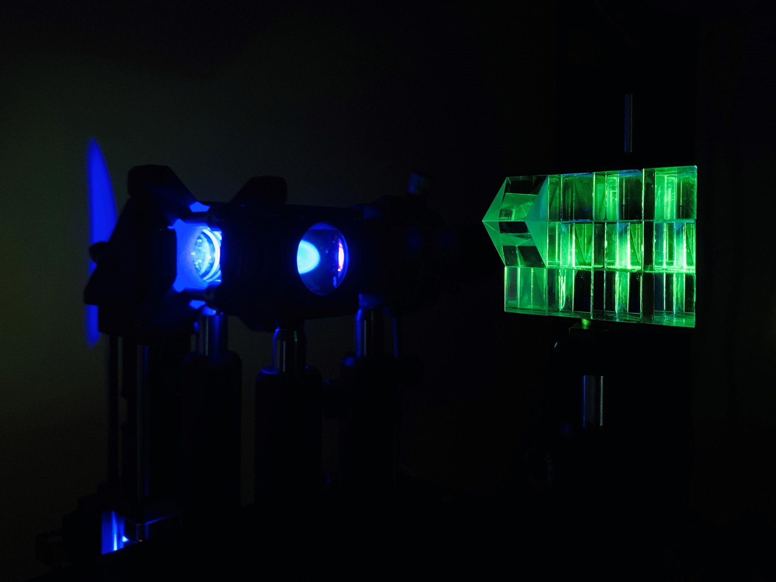
The researchers developed a new multifocus technique that uses a Z-splitter prism (right) to split detected light in a standard microscope. This simultaneously creates multiple images, each focused at a different depth in the sample, in a single camera frame. Photo credit: Sheng Xiao, Boston University
The new approach captures multi-focus microscope images at high speed.
Researchers have developed a simple method to simultaneously acquire images at different depths with a standard microscope. The new technique can be applied to a variety of microscopy methods, making it useful for a variety of biological and biomedical imaging applications.
“Optical microscopy has been an indispensable tool for studying complex 3D biological systems and processes,” said Sheng Xiao, a member of the Boston University research team. “With our new multifocus technology, living cells and organisms can be observed at high speed and high contrast.”
in the OpticaResearchers from the Optical Society (OSA) for Highly Effective Research, led by Jerome Mertz, describe their new simple and fast way to get information from different depths using standard microscopy. The new approach can be easily added to most existing systems and is easy to replicate so that it is accessible to other researchers.
Taking multi-focus images
Standard camera-based microscopy systems capture sharp images in a single focal plane. Although researchers have tried different strategies to simultaneously capture images with different focal lengths, these approaches typically require multiple cameras or use a special diffractive optical element to perform the image division with a single camera. Both strategies are complex and it can be difficult to make a diffractive optical element.
“We used a Z-splitter prism that can be assembled entirely from off-the-shelf components and easily applied to a variety of imaging modalities such as fluorescence, phase contrast, or dark field imaging,” said Xiao.
The Z-splitter prism splits detected light to produce multiple images simultaneously in a single camera frame. Each image is focused to a different depth in the sample. Using a high-speed camera with a large sensor area and a high number of pixels allowed the researchers to distribute multiple high-resolution images without overlapping on the same sensor.
The multifocal images recorded with the new technology make it possible to estimate the blurred background from the sample much more precisely than is possible with a single image. The researchers used this information to develop an improved 3D deburring algorithm that eliminates the blurry background light that is often a problem when using wide-field microscopy.
“Our 3D enhanced volume deblurring algorithm suppresses blurred background from sources outside the image volume,” said Xiao. “This improves both the image contrast and the signal-to-noise ratio and is therefore particularly advantageous in fluorescence imaging applications with thick samples.”
Proven versatility
Researchers demonstrated the new technique using commonly used microscopy modalities, including fluorescence, phase contrast, and dark field imaging. They took large field-of-view 3D images that spanned hundreds of neurons or entire freely moving organisms, as well as high-speed 3D images of a rotifer cilia beating every hundredth of a second. This demonstrated how the approach offers the flexibility to prioritize a large field of view or high speed.
To demonstrate the capabilities of the 3D deblurring algorithm with expanded volume, the researchers imaged various thick samples, including the brain of a living mouse. They observed significant improvements in contrast and signal-to-noise ratio compared to raw multifocus images and more traditional 3D deburring algorithms. The researchers are now working to expand the technology so that it works with even more imaging modalities.
Reference: “High-contrast multifocus microscopy with a single camera and a Z-splitter prism” by Sheng Xiao, Howard Gritton, Hua-An Tseng, Dana Zemel, Xue Han and Jerome Mertz, October 22, 2020, Optica.
DOI: