The thin and inexpensive lens is 3D printed and can examine living cells, which would greatly improve the diagnosis. Photo credits: © 2020 KAUST; Andrea Bertoncini
Customer-specific lenses make it easier to attach high-tech microscopes directly to cell incubators.
An optical device similar to a miniaturized lighthouse lens can make it easier to look into Petri dishes and observe details of biological processes at the molecular level, including the growth of cancer cells. The new lens developed by KAUST is also very inexpensive.
Many bioimaging techniques require the addition of fluorescent dyes to specific cell targets. However, a recently developed method known as Stimulated Raman Scattering (SRS) microscopy can avoid cumbersome labeling steps by using laser pulses to collect molecular vibrational signals from biological samples. The ability of SRS microscopes to produce high-resolution, non-invasive images in real time has led researchers to use them for too in vivo disease diagnostic studies.
KAUST researchers have developed an ultra-thin lens that fits into the on-stage incubators that are used to cultivate living cells for bioimaging. Photo credit: © 2020 KAUST
A disadvantage of SRS microscopes, however, is that the detection system is affected by a background signal known as cross-phase modulation, which is generated by the intense interactions between laser pulses and the samples.
“This background signal is omnipresent and reduces the contrast when observing complex samples such as living cells under the microscope,” explains Carlo Liberale from KAUST. “It also makes it difficult to identify target molecules.”
To avoid the effects of cross-phase modulation, most SRS microscopes must use bulky glass lenses that can capture large angles of light. However, it is next to impossible to fit these types of lenses into the on-stage incubators that are used to grow live cells for bioimaging.
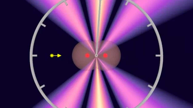
A 3D printed lens developed at KAUST uses optical features inspired by lighthouse rays to collect laser signals for bioimaging. Photo credit: © 2020 Andrea Bertoncini
Andrea Bertoncini, a researcher in Liberales Group, pioneered the development of an ultra-thin SRS lens with laser-based three-dimensional (3D) printing. The KAUST team took its cue from the slim design of lighthouse lenses and printed tiny lens and mirror-like features in a transparent polymer that was only a fraction of a millimeter thick.
“This type of lens design is a very efficient way of collecting and redirecting light from wide-angle sources directly to our laser detector,” says Bertoncini. “And because it’s so thin, it fits easily into the closed chambers of an incubator.”
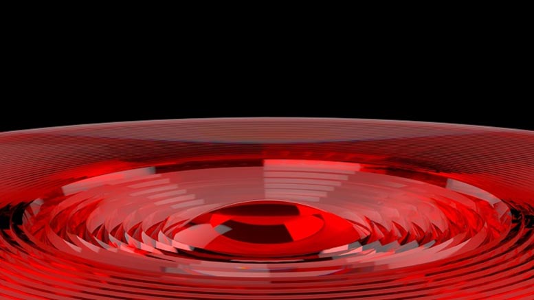
After calibration trials confirmed that their new lens could reject the background of cross-phase modulation, the researchers turned their attention to human cancer cells that were cultured in a conventional Petri dish. These experiments showed that the lens was able to image the internal components of the cell with a resolution similar to traditional SRS microscopes, but in a much more convenient and inexpensive format.
“The targets we normally use to collect SRS microscope signals cost a few thousand dollars,” says Bertoncini. “Now we have a lens with similar advantages that we can make for less than a tenth that price.”
Reference: “3D-printed catadioptric thin lens with high NA to suppress the XPM background in stimulated Raman scattering microscopy” by Andrea Bertoncini, Sergey P. Laptenok, Luca Genchi, Vijayakumar P. Rajamanickam and Carlo Liberale, July 13, 2020, Journal of Biophotonics.
DOI: 10.1002 / jbio.202000219