
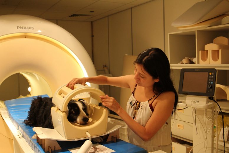
Researchers from the Department of Ethology at Eötvös Loránd University trained the dogs to scan the brains of alert, unbridled dogs. Photo credit: Eniko Kubinyi / Eötvös Loránd University
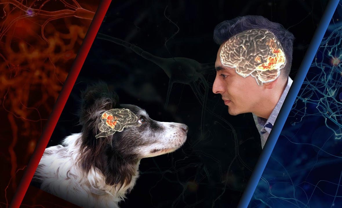
Scientists discover remarkable similarities and differences in the way the brains of dogs and humans process visual information about others.
Researchers at the Institute of Ethology at Eötvös Loránd University in Hungary discovered remarkable similarities and differences in the way dogs and human brains process visual information about others. The study was published in The Journal of Neuroscience on October 5, 2020.
Faces are central to visual communication in humans, who have a special neural network for face processing. Although dogs also pay attention to faces, are distinguished by eye contact and reading facial emotions, they also rely on additional body signals to communicate. Like human brains, do dog brains specialize in face processing?
To investigate similarities and differences in how the dog and human brain respond to visual information about others, the researchers tested twenty dogs and thirty humans in the same functional magnetic resonance imaging (fMRI) experiment. Dogs and humans watched short films of dogs and human faces and to compare dogs and human heads. Specifically, this research is the first in-line, non-invasive, visual neuroimaging study of a non-primate and a primate species.
Video summary of the research:
The study was carried out in several locations: researchers have joined forces from two of the few laboratories in the world capable of scanning the brains of awake, unbridled dogs (Institute of Ethology, Faculty of Science, Eötvös Loránd University, Budapest, Hungary and Institute of Neurobiology), National Autonomous University of Mexico, Querétaro, Mexico) to collect brain response data from more dogs than has been done in most canine fMRI studies to date.
In terms of similarities, the study identified areas of the brain in both dogs and humans that reacted differently to videos depending on whether they showed an individual of their own species. “Our research group has previously shown a similar correspondence between the brains of dogs and humans for language processing. We now see that species sensitivity is an important organizing principle in the mammalian brain to process social stimuli in both auditory and visual modality ”- explains Attila Andics, lead author of the study.
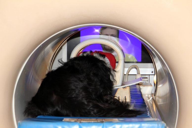
Dogs and humans watched short films of dogs and human faces and to compare dogs and human heads. Photo credit: Eniko Kubinyi / Eötvös Loránd University
In terms of differences, the study did not find any areas of the brain in dogs that encode whether the image being viewed is a face or the back of the head – while this is a crucial distinction in humans. “A preference analysis of the brain reaction patterns confirmed that in dogs the preference for conspecifics over the preference for the face and in humans the preference for the face over the preference for conspecifics. This is a major difference. It shows that cortical specialization for facial perception can vary significantly in mammals. In fact, these results also shed new light on previous canine fMRI studies that claimed they found “facial areas”: We now believe that the increased activity on canine faces in these studies suggests that brain areas are more likely than dogs to prefer Prefer faces ”- explains Nóra Bunford, joint first author of the study and coordinator of data collection in Hungary.
Dogs are trained to remain still during the fMRI exam. Photo credit: Eniko Kubinyi / Eötvös Loránd University
The researchers also identified regions of the canine and human brain that showed a similar pattern of activity in response to the videos. “This so-called representative similarity analysis can directly compare brain activity patterns between species. Interestingly, the similarities between dog and human activity patterns were stronger for what we called functional matching (comparing the activity for the dog’s face in the dog’s brain with the activity for the human face in the human brain) than for physical matching (comparing the Activity for dog face in dog brain) dog brain for activity for dog face in human brain). This shows that we may have resorted to high-level categorical processing of social information rather than low-level visual processing in both dogs and humans “- explains Raúl Hernández-Pérez, the other lead author of the study and coordinator of data collection in Mexico.
“Taken together, the similarities in species sensitivity and the differences in facial sensitivity suggest both functional analogies and differences in the organizational principles of visuosocial processing between dogs and humans. This is another demonstration that comparative imaging with phylogenetically distant mammalian species can improve our understanding of how social brain functions are organized and how they have evolved, ”Andics concludes.
Reference: “Comparative brain imaging shows analogous and different patterns of species and facial sensitivity in humans and dogs” by Nóra Bunford, Raúl Hernández-Pérez, Eszter Borbála Farkas, Laura V. Cuaya, Dóra Szabó, Ádám György Szabó, Márta, Ádám Miklósi and Attila Andics, October 5, 2020, The Journal of Neuroscience.
DOI: 10.1523 / JNEUROSCI.2800-19.2020
This study was published in The Journal of Neuroscience The title “Comparative Brain Imaging Shows Analog and Different Patterns of Species and Facial Sensitivity in Humans and Dogs”, written by Nóra Bunford, Raúl Hernández-Perez, Eszter Farkas, Laura Cuaya, Dóra Szabó, Ádám Szabó, Márta Gácsi, Ádám Miklósi and Attila Andics. The study and researchers were funded by the Hungarian Academy of Sciences (Lendület Program LP2017-13 / 2017, LP2018-3 / 2018; and F01 / 031), the Mexican National Council for Science and Technology (CONACYT; 409258 and 407590) . the National Bureau for Research, Development and Innovation (115862K), the Hungarian Ministry of Human Resources (ÚNKP-17-4-ELTE / 12423/11), the European Research Council (ERC; 680040), the Eötvös Loránd University (ELTE) and the National Brain Research Program (2017-1.2.1-NKP-2017-00002).