
A new approach called integrated neurophotonics could allow researchers to track the activity of all the neurons that make up a given brain circuit. Photo credit: Roukes et. al.
A new approach called integrated neurophotonics could allow researchers to track the activity of all the neurons that make up a given brain circuit.
In order to deepen their understanding of the brain, neuroscientists need to be able to detail the neural circuits that are responsible for tasks such as processing sensory information or creating new memories. Now a team of Caltech researchers has described a new approach with which the activity of thousands to millions of neurons in a given brain circuit can be observed in real time. The novel method discussed in an article published in the journal “Perspective” Neuron on October 14, 2020 has far greater potential than any current approach, say the authors.
The new technique, known as “integrated neurophotonics”, uses tiny arrays of optical microchips that, in combination with fluorescent molecular reporters and optogenetic actuators, can be implanted at any depth of the brain to optically monitor neurons and control their activity. The arrays send out microscale beams of light to stimulate the genetically modified neurons around them while recording the activity of these cells and revealing their function. Although the work is currently only done in animal models, it could one day help unravel circuits deep in the human brain, says Michael Roukes, principal researcher on the paper and Frank J. Roshek professor of physics, applied physics and bioengineering at Caltech.
“Dense uptake in depth – that’s the key,” says Roukes. “We will not be able to record all the activity of the brain anytime soon. But could we focus on some of its important computational structures within certain brain regions? That is our motivation. ”
In recent years, neuroscientists have begun using optogenetics to study ever larger groups of neurons in model animals, including rodents. In optogenetics, neurons are genetically modified in such a way that they express a certain protein marker such as green fluorescent protein (GFP) when they are excited by a certain wavelength of light. The presence of GFP causes the cell to glow green under fluorescent light, which is a visual indicator of neural activity. By fusing sensor molecules with these markers, researchers can construct neurons that signal their local activity by modulating this fluorescence. Optogenetics solves some problems inherent in neuroscientific studies that use implanted electrodes to measure the electrical activity of neurons that, on average, can only reliably measure a single neuron due to the total electrical activity in the brain. Because the brain doesn’t use light to communicate, optogenetics makes it easy to track a large number of these neural signals.
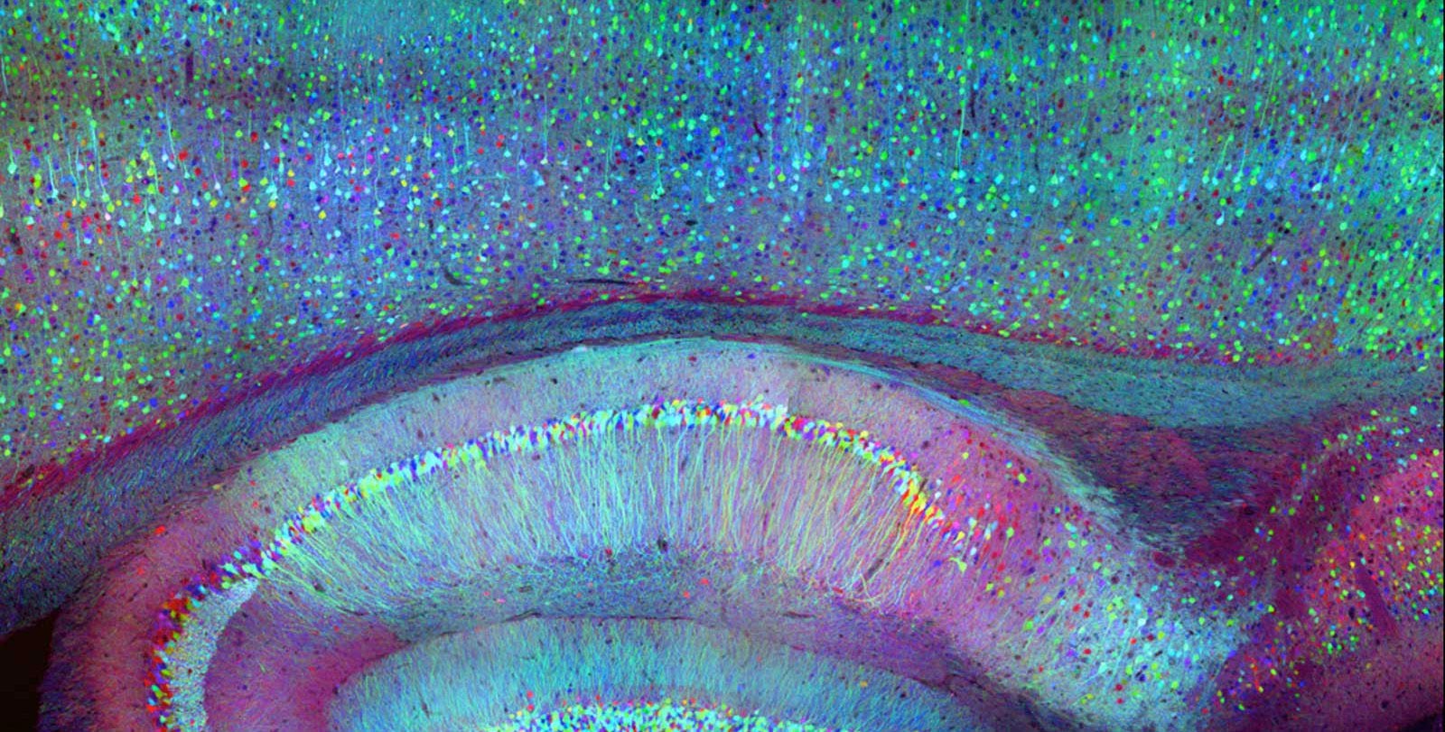
Current optical techniques can only map the activity of neurons near the surface of the brain, but integrated neurophotonics could unlock circuits buried deep in the brain. Photo credit: Roukes et. al.
However, current optogenetic studies of the brain are limited by a significant physical limitation, says Laurent Moreaux, senior research scientist and lead author at Caltech. Brain tissue scatters light, which means that light entering from outside the brain can only travel short distances within the brain. For this reason, only regions that are less than two millimeters from the surface of the brain can be examined optically. Because of this, the best-studied brain circuits are usually simple circuits that relay sensory information, like a mouse’s sensory cortex – they’re located near the surface. In short, at this time, optogenetic methods cannot readily provide insight into circuits deeper in the brain, including those involved in cognitive or higher-order learning processes.
Integrated neurophotonics, according to Roukes and colleagues, avoids the problem. In this technique, the microscale elements of a complete imaging system are implanted near complex neural circuitry deep within the brain, in regions such as the hippocampus (which is involved in memory formation), the striatum (which controls perception), and other basic ones Structures in unprecedented resolution. Consider the similar technology of functional magnetic resonance imaging (fMRI), the scanning technique currently used to image whole brains. Each voxel, or three-dimensional pixel, in an fMRI scan is typically about one cubic millimeter in volume and contains about 100,000 neurons. Each voxel therefore represents the average activity of all of those 100,000 cells.
“The overall goal of integrated neurophotonics is to record what everyone The neuron in this collection of 100,000 works in real time, ”says Roukes.
Roukes’ long-term goal is to disseminate the advanced instrumentation of integrated neurophotonics to enable multi-institutional collaborations that will drive advanced neuroscientific research using this novel technology. Previously, he says, this type of neurotechnology development relied mostly on research led by an individual laboratory or researcher. Starting in 2011, Roukes worked with five other scientists and the White House Science and Technology Policy Bureau to launch the United States’ BRAIN (Brain Research Through Fostering Innovative Neurotechnologies) initiative, launched during the Obama administration. Her vision was to bring into neuroscientific research the kind of large-scale partnerships seen in the natural sciences, such as hardware development projects such as international telescope collaborations and the LIGO-Virgo to find cooperation Gravitational waves. According to Roukes, integrated neurophotonics opens doors for such teamwork in instrument making
“Lots of the building blocks [for an approach such as ours] has been around for a decade or more, ”he says. “But until recently, there just wasn’t the vision, will, and resources to bring them all together to realize these powerful new tools for neuroscience.”
The paper describing this research is entitled “Integrated Neurophotonics: Towards a Dense Volumetric Inquiry into Cerebral Circulatory Activity – In Depth and Real Time”. Other Caltech co-authors are Wesley D. Sacher, a former postdoc at the Kavli Nanoscience Institute, and former Caltech postdoc Nicole J. Kubat. The work, which involved staff from 14 other institutions, was funded by a grant from the National Institutes of Health’s BRAIN Initiative, the Agency for Advanced Defense Research Projects, the National Science Foundation, and the Kavli Foundation.
Reference: “Integrated Neurophotonics: Towards a Dense Volumetric Query of the Activity of the Brain Circulatory System – in Depth and in Real Time” by Laurent C. Moreaux, Dimitri Yatsenko, Wesley D. Sacher, Jaebin Choi, Changhyuk Lee, Nicole J. Kubat and R. James Cotton, Edward S. Boyden, Michael Z. Lin, Lin Tian, Andreas S. Tolias, Joyce KS Poon, Kenneth L. Shepard and Michael L. Roukes, October 14, 2020, Neuron.
DOI: 10.1016 / j.neuron.2020.09.043
CaltechAUTHORS: 20201014-111855866